Fig. 3.
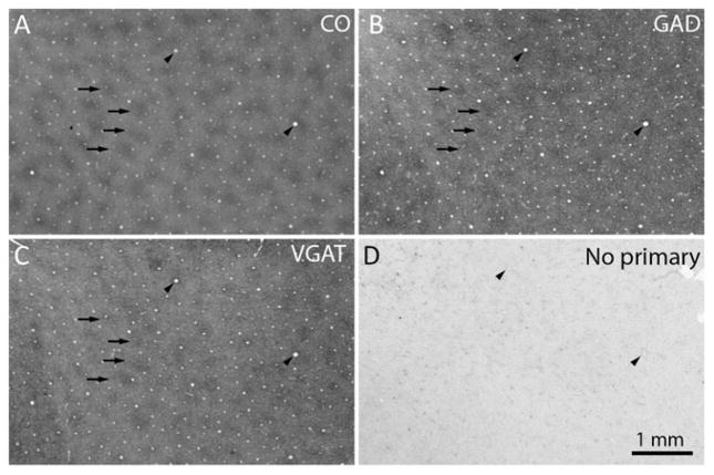
GAD and VGAT are localized preferentially in CO patches. (a) CO patches, with 4 examples highlighted by arrows. The arrowheads mark prominent blood vessels, which allow precise alignment of adjacent sections. (b) GAD patches, which match the CO patches (arrows). (c) VGAT patches, which match the CO and GAD patches (arrows). (d) Control section omitting the primary antibody shows only light background labeling from endogenous peroxidase activity in red blood cells.

