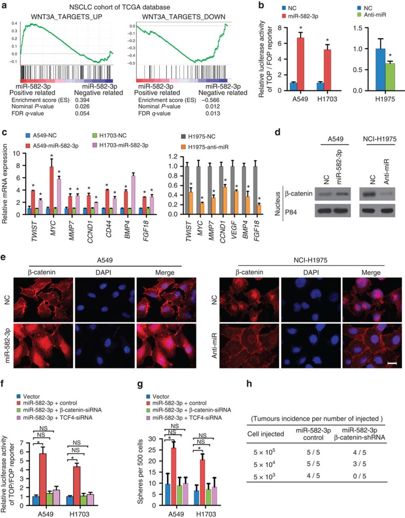
Figure 5. miR-582-3p activates Wnt/β-catenin signalling.
(a) GSEA plot showing that miR-582-3p expression was positively correlated with Wnt-activated gene signatures and inversely correlated with Wnt-suppressed gene signatures in the TCGA lung cancer data set. (b) The indicated cells were transfected with TOP or FOP reporter and Renilla pRL-TK plasmids and subjected to dual-luciferase assays 48 h after transfection. The detected reporter activity was normalized to the Renilla activity. (c) RT–qPCR analysis of the expression of the established downstream targets for the Wnt/β-catenin pathway, including TWIST, MYC, MMP7, CCND1, CD44, BMP4 and FGF18, in the indicated cells. (d) Altered nuclear translocation of β-catenin in response to ectopic miR-582-3p expression. Nuclear fractions of the indicated cells were analysed by western blot analysis. P84 was used as a loading control. (e) Subcellular β-catenin localization in the indicated cells was assessed by immunofluorescence staining. Scale bar, 20 μm. (f) Luciferase assay of TCF/LEF transcriptional activity in indicated cells. (g) Representative images and quantification of cellular spheres formed by the indicated cells. (h) Tumour formation frequencies for the different numbers of indicated cells. Each bar represents the mean±s.e.m. derived from three independent experiments. A two-tailed Student's t-test was used for statistical analysis (*P<0.05, NS: not statistically significant).

