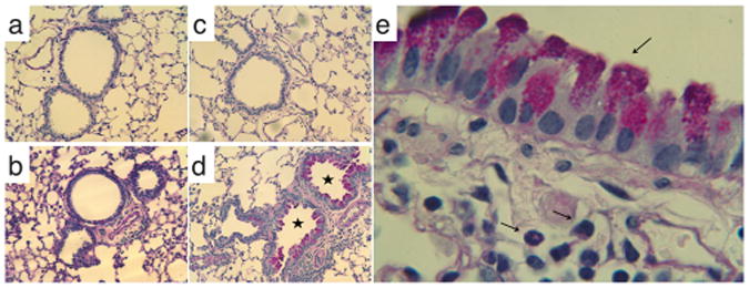
Figure 2.

Lung tissue inflammation and mucus production evoked by GSH–MDI reaction products. Lung tissue sections from naïve mice exposed to control stimuli, GSH-m (a), or GSH–MDI (b) or from MDI sensitized mice exposed to control stimuli, MDI-m (c), or GSH– MDI (d) were subject to periodic acid–Schiff (PAS) staining to highlight mucus production (airways with asterisk). (e) PAS stained lung tissue section from a representative MDI sensitized, GSH–MDI exposed host under higher magnification to highlight the mucus containing goblet cells lining the airways and submucosal eosinophils.
