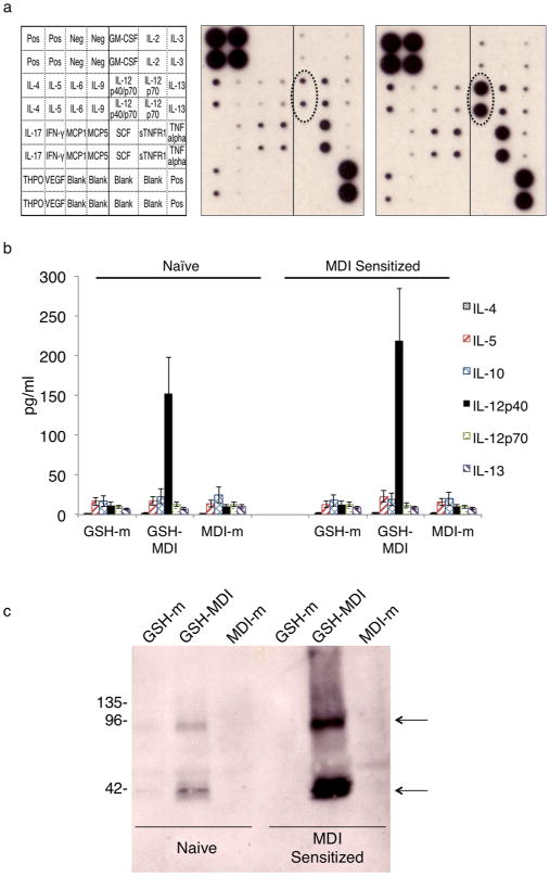
Figure 3.

GSH–MDI increases airway levels of IL-12/IL-23β. (a) Monoclonal antibody-based array (key to the left) was used to screen for changes in cytokine levels of pooled airway fluid samples (N = 6 each) from MDI sensitized hosts exposed to control stimuli MDI-m (MDI reacted without GSH) vs GSH–MDI (middle and far right respectively). (b) Graph representing the mean concentration in pg/ mL ± standard error of different cytokines in airway fluid (Y-axis) from N = 18 each naïve or MDI sensitized mice from three separate experiments, exposed to GSH–MDI or control stimuli, as labeled. (c) Anti-IL-12/IL-23β western blot on airway fluid from naïve or MDI sensitized mice exposed to GSH–MDI or control stimuli (GSH-m = GSH reacted without MDI; MDI-m = MDI reacted without GSH) was performed under nonreducing conditions. Arrows highlight banding due to monomeric p40 (lower) and homodimeric p80 (upper) IL-12/ IL-23β.
