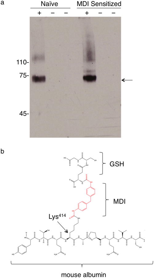
Figure 5.

Modification of airway fluid albumin by GSH–MDI in vivo. (a) Pooled airway fluid samples from N = 6 each naïve or MDI sensitized mice exposed to GSH–MDI (+) or control stimuli (−), GSH-m and MDI-m, respectively, were subject to reducing SDS-PAGE and western blotted with biotin labeled MDI-specific mAb DA5. Arrow highlights dominant band ∼68 kDa. (b) Chemical structure depicting the unique GSH–MDI modification of airway fluid albumin detected through LC-MS/MS (see Supporting Information Tables S1 and S2).
