
Figure 1.
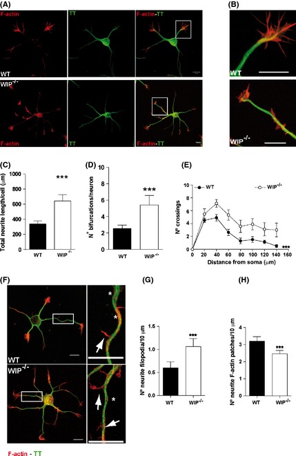
WIP deficiency increases neuritic branching and filopodium formation in dissociated hippocampal murine neurons. (A) Representative confocal images of dissociated embryonic WT and WIP −/− hippocampal neurons (E18) fixed 24 h post plating and stained for F‐actin (TRITC‐phalloidin, red) and MT (Alexa488‐anti‐tyrosinated tubulin (TT), green). WIP −/− neuron morphology was more complex than that of WT neurons. Scale bar, 10 μm. (B) High magnification images of the insets in A. Scale bar, 10 μm. (C) Average total neuritic length increased in 24‐h‐cultured WIP −/− compared to WT hippocampal neurons. The morphology of 30 neurons per genotype was characterized. (D) Average number of bifurcations per neuron increased in WIP −/−compared to WT neurons. (E) Sholl analysis of traced WT and WIP −/− neurons showing higher number of crossings in WIP −/− neurons. n = 17 neurons analyzed per experimental group, from three independent experiments. ***P < 0.001 (Two‐way ANOVA). (F) Representative confocal images of WT and WIP −/− neurons obtained and stained as in (A). Right images show higher magnification of the boxed area in left images (scale bar, 10 μm for both). Neuritic filopodia (arrows), neuritic actin patches (asterisks). (G,H) Quantification of neuritic filopodia and actin patches per 10 μm in WT and WIP −/− neurons. Data for 50 neurons per group in three independent experiments. Student's t‐test; ***P < 0.001.