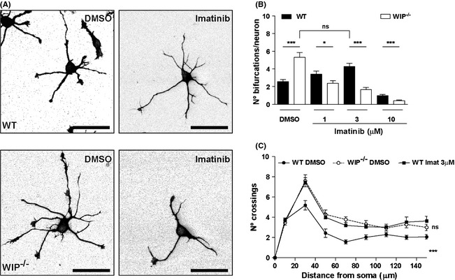
Figure 4.

Imatinib inhibition of Abl kinases increases neuritic bifurcations in WT cells, mimicking WIP deficiency. (A) Representative images of dissociated WT and WIP −/− hippocampal neurons incubated for 24 h with DMSO or 3 μM imatinib, fixed, and stained with anti‐TT mAb. Scale bar, 50 μm. (B) Quantification of experiments in (A). Note the larger number of bifurcations in WIP −/− neurons in DMSO, which is equivalent to that of WT neurons in 3 μM imatinib. n = 150 neurons analyzed per experimental group, from three independent experiments; ns, not significant; *P < 0.05; ***P < 0.001 (Student's t‐test). (C) Sholl analysis of traced neurons, after imatinib treatment of WT and WIP −/− cells. The quantification shows higher number of crossings in WIP −/− neurons compared to WT cells before treatment. After treatment with imatinib 3 μM, WT neurons show increased branching compared to DMSO‐treated WT neurons. n = 30 neurons analyzed per experimental group, from three independent experiments. ***P < 0.001 (WIP −/− neurons compared to WT cells before treatment); ns, not significant (imatinib‐treated WT neurons compared to DMSO‐treated WIP −/− cells) (Two‐way ANOVA).
