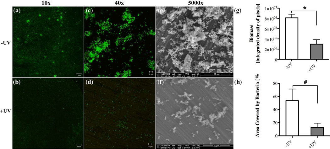
Fig. 6.
Effect of UV-treatment of titanium surfaces on bacterial biofilm formation after 16 hours incubation evaluated via confocal microscopy imaging through (a,b) 10x and (c,d) 40x objectives with representative images illustrating the live/dead distribution of bacterial cells (green for live cells, red for compromised cells) accumulated on (a,c) untreated (-UV) and (b,d) UV-treated (+UV) titanium discs. Scanning electron microscopy revealing the biofilm structure on (e) -UV and (f) +UV titanium discs. Quantitative comparison of (e) accumulated biomass and (f) the area covered by bacteria between untreated (white bar, –UV) in comparison to UV-treated (gray bar, +UV) titanium discs. Each value represents the mean ± SD of four samples comprised of two technical replicates of two independent biological experiments. Statistically significant differences are indicated as: * p<0.0116), # p<0.0461.

