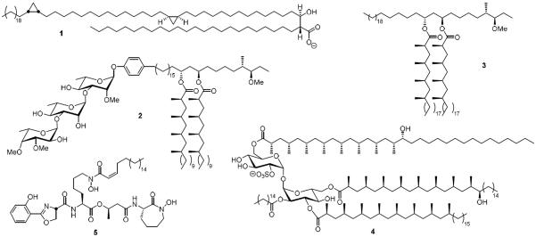
Figure 1.
Unique lipids and lipidated metabolites found in cell envelope of Mycobacterium tuberculosis. All of the molecules shown exist as a suite of related isomers that vary in the lipid chain length. If reported, the major isomer is shown otherwise a representative molecule is depicted. The mycolic acids are represented by the most abundant a mycolic acid (α-MA, 1), the phenolic glycolipids are represented by 2, the phthiocerol dimycocerosates are exemplified by PDIM-A (3), the sulfolipids are represented by SL-1 (4) and the mycobactins by 5.

