
Figure 2.
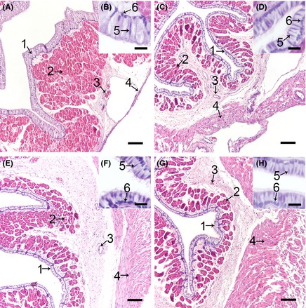
The Histological Structure of the Turtle Oviduct after H&E Staining. Cross‐sections of the magnum (A), isthmus (C), uterus (E), and vagina (G) of the oviduct, and the amplification of the epithelium in the magnum (B), isthmus (D), uterus (F), and vagina (H). Every part showed a similar structure. Epithelial cell layer (1), secretory gland (2) in the lamina propria, and blood vessels (3) in the submucous and muscle layers (4) are shown. In the epithelial cell layer, two cells types were observed: ciliated cell nucleus (5) and secretory cell nucleus (6). Scale bars: 100 μm (A, D, E, and G) and 20 μm (B, D, F, and H).