
Figure 3.
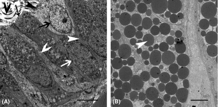
The Ultrastructure of the Epithelium and Secretory Glands. (A) Transverse sections throughout the epithelial cell; the ciliated cells have cilium (black fat arrow), which are located on the ciliated cell superior surface (black thin arrow), but not on the secretory cell superior surface (black thin arrow). The ciliated cell nucleus (black fat arrow) was similar in size to the secretory cell nucleus (white fat arrow). The junction (white thin arrow) between two secretory cells was observed. The junction (white fat arrow) between the secretory cell and the ciliated cell was also observed. We also observed the secretory cell basic membrane (white thin arrow). (B) Transverse sections throughout the secretory gland. The secretory glands were filled with many secretory particles (white thin arrow). Scale bar: 5 µm (A and B).