
Figure 6.
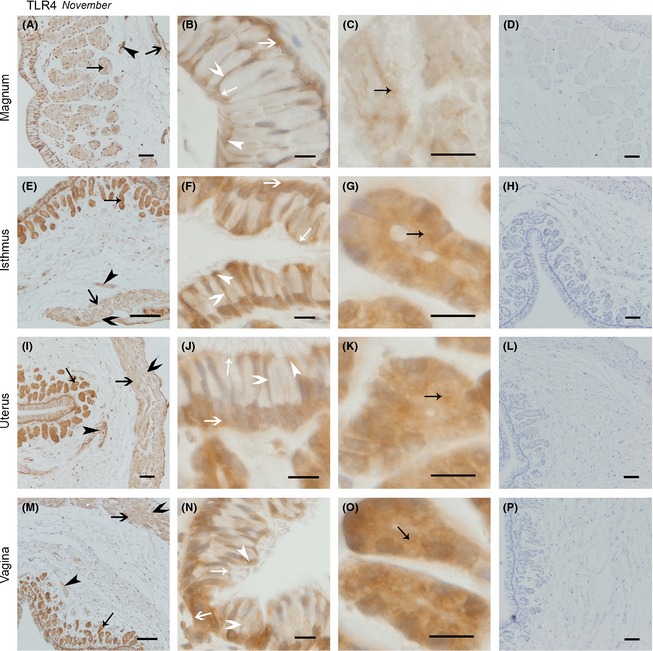
The Expression of TLR4 protein in November was Similar in the Magnum, Isthmus, Uterus, and Vagina. Cross‐sections of the magnum, isthmus, uterus, and vagina are shown at low magnification in A, E, I, and M, respectively; B, F, J, and n refer to the amplification of the epithelium in the magnum, isthmus, uterus, and vagina, respectively; the secretory glands in magnum, isthmus, uterus, and vagina are described in C, G, K, and o; respectively; and D, H, I, and P represent the negative controls for the magnum, isthmus, uterus, and vagina, respectively. No immunoreaction products were observed. All sections were counterstained with hematoxylin. Positive staining was observed on the ciliated cell superior surface and cilium surface (white thin arrow head), secretory cell superior surface (white thin arrow), secretory cell lateral membrane (white fat arrow head), secretory cell basal membrane (white fat arrow), secretory gland vesicles membrane (black thin arrow), longitudinal muscle (black fat arrow), circular muscle (black fat arrow head), blood vessel endothelium (black thin arrow head). Scale bars: 100 μm (E and M), 50 μm (A, D, H, I, L, and P), and 10 μm (B, C, F, G, J, K, N, and O).