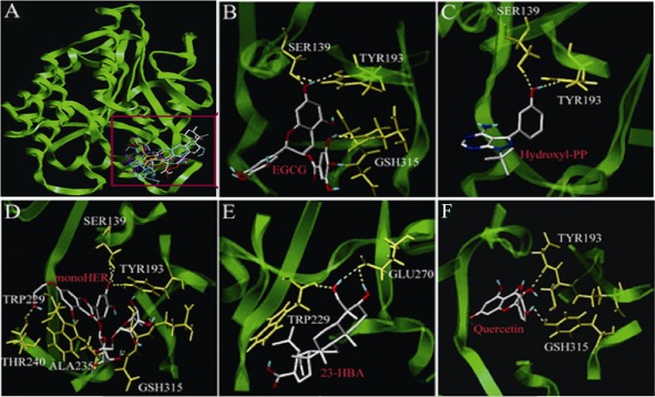
Figure 8.

Molecular docking analysis of 23‐HBA and four other CBR1 inhibitors towards CBR1 protein. (A) Three‐dimensional model of the active binding pocket of polymorphic human CBR1 with 23‐HBA and other inhibitors. CBR1 (green) is shown in ribbon form. 23‐HBA (grey), quercetin (red), MonoHER (blue), hydroxy‐PP (yellow) and EGCG (purple) are represented in stick form. (B) Active site of human CBR1 with EGCG. (C) Active site of human CBR1 with hydroxyl‐PP. (D) Active site of human CBR1 with monoHER. (E) Active site of human CBR1 with 23‐HBA. (F) Active site of human CBR1 with quercetin.
