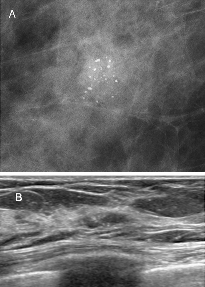
Fig 1. A 64-year-old asymptomatic female with an abnormality on screening mammography.
(A) Magnification mammography demonstrates pleomorphic clustered microcalcifications in the right lower outer breast. (B) This lesion shows tubular isoechoic lesion with microcalcifications on US. The pathologic findings of both Wi-UVAB and surgical excision were pleomorphic LCIS.

