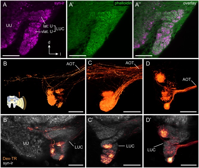
Fig 3. Transmedulla neurons: Ramifications within the lower unit complex of the anterior optic tubercle (AOTU-LUC).
(A) Confocal image of synapsin-ir (syn-ir, magenta) and f-actin labeling (phalloidin, green) of the anterior optic tubercle. The AOTU-LUC has been previously divided into the lateral unit (lat. U) and the ventrolateral unit (vlat. U) [55]. Synapsin/phalloidin labeling reveals that these two units are further structured into a complicated assembly of multiple small subcompartments. B-D) Direct volume rendering of AOTU-LUC projections from transmedulla neurons labeled through dextran Texas Red (Dex-TR) injection into the MEDRA. In each sample a different combination of focal projection areas is stained. Cartoon in (B) illustrates injection site. B’-D’) Single confocal sections (B’, C’) and maximum intensity projection of 10 adjacent slices (D’) of the neurons shown in B-D, combined with anti-synapsin labeling (grey). Projections from MEDRA neurons are exclusively found in the AOTU-LUC. AOT, anterior optic tract; UU, upper unit of the anterior optic tubercle. All views in frontal plane. All scale bars: 30 μm.

