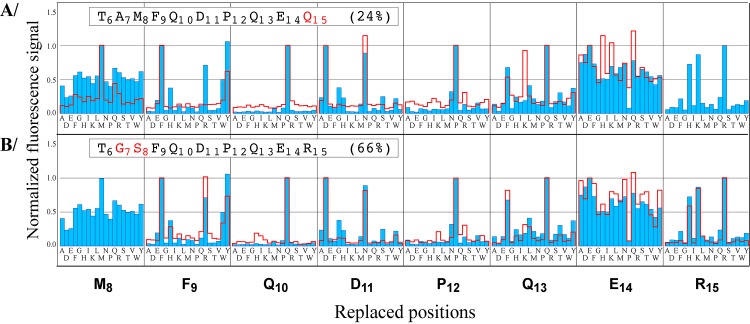
Fig 3. WT-like permutation patterns (positions 8–15).
The patterns of variants R15Q (A) and A7G-M8S (B) are represented as red lines and superimposed with the WT pattern (blue). Fluorescence signals were normalized with respect to that recorded with each starting peptide. Patterns at replaced positions are not shown because they are not relevant to the modified sequence context.

