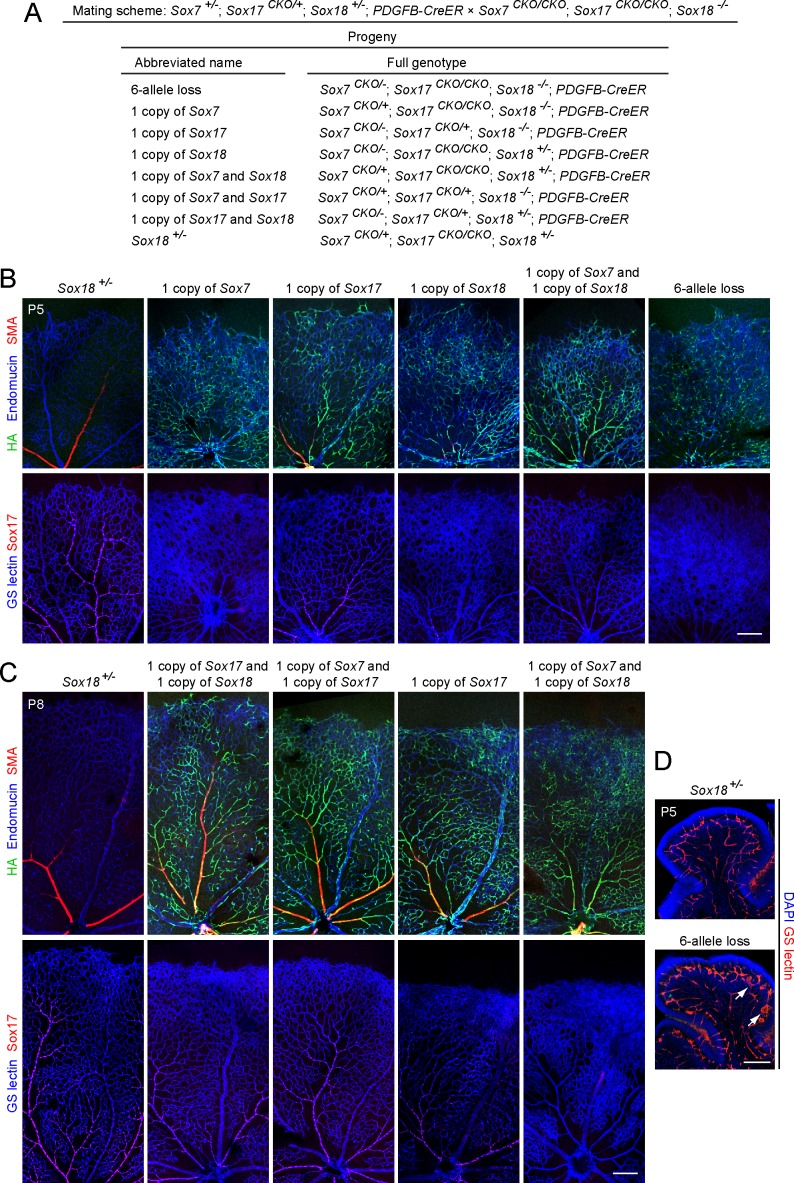
Fig 4. Combined loss of Sox7, Sox17, and Sox18 in ECs alters retinal vasculature.
(A) Summary of the triple SoxF cross and the resulting progeny with abbreviations for their genotypes. (B, C) Analysis of retinal vasculature in SoxF mutants at P5 (B) and P8 (C). Top panels, retina flat mounts immunostained with anti-endomucin, anti-SMA and anti-HA. Bottom panels, retina flat mounts stained with GS-lectin and anti-Sox17. Anti-HA and anti-Sox17 staining assesses CreER-mediated recombination efficiency. The retinal vasculature in Sox18 +/- is indistinguishable from WT. The mutants that lack Sox17 and either one or both copies of Sox7 and Sox18 show greatly reduced vSMC coverage of radial arteries with increased capillary density. 50–100 μg 4HT was given at P0 and P2. Scale bar, 200 μm. (D) Cerebellum sections from Sox7 CKO/- ;Sox17 CKO/CKO ;Sox18 -/- ;Pdgfb-CreER and Sox18 +/- control at P5, following 50–100 μg 4HT at P0 and P2. Vascular disorganization (arrows) is seen in Sox7 CKO/- ;Sox17 CKO/CKO ;Sox18 -/- ;Pdgfb-CreER. Scale bar, 200 μm.

