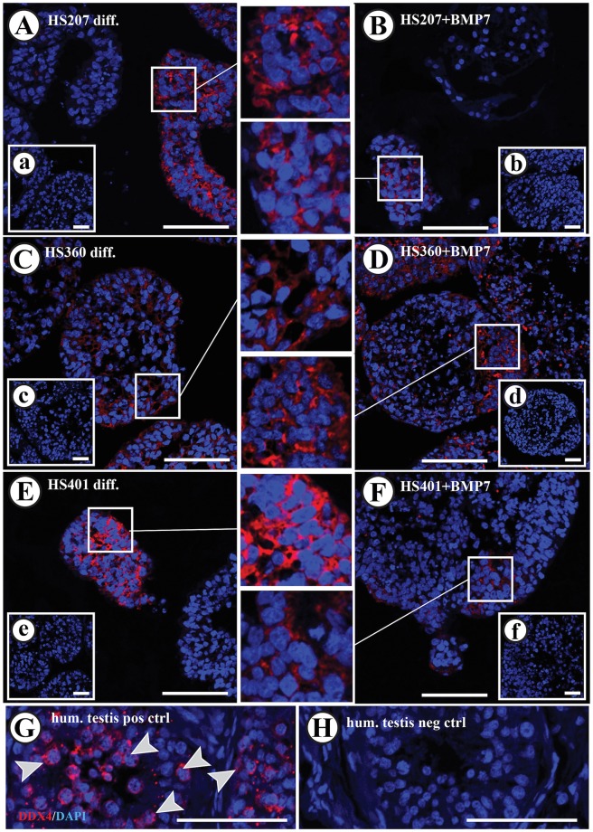
Fig 6. Immunofluorescence detection of DDX4 in HS207, HS360 and HS401 cells cultured in suspension.
Cytoplasmic expression of DDX4 was observed in all hES cell lines after spontaneous differentiation (A, C and E) and after stimulation with BMP7 (B, D and F). Cytoplasmic expression of DDX4 was observed in germ cells present in adult human testis (G; arrow heads). Negative controls exhibited no specific staining in either the nucleus or cytoplasm (H and small inserts in A to F). Red: DDX4 staining. Blue: DAPI staining marking the nucleus. Scale bars: 100 μm in A to H, and 50 μm in the small inserts.

