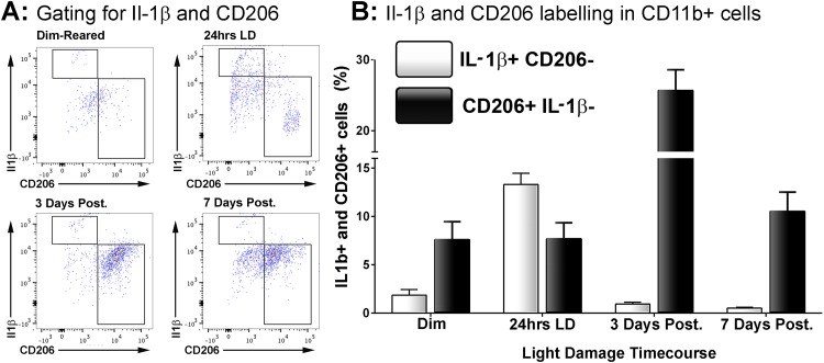
Fig 3. Correlation of CD206 and Il-1β immunolabelling within the CD11b+ macrophage population following LD.
A: Representative flow cytometry plots examine CD206+ and Il1b+ cell counts within the CD11b population following light damage. For the most part, Il-1β and CD206 cells occupied mutually distinct subsets within the population CD11b cells. B: Quantification of Il-1β+/CD206- and CD206+/Il-1β- cells as percentage of the CD11b+ population following LD. There was a sharp increase in the proportion of Il-1β+/CD206- cells immediately following 24hrs LD (P<0.05), though this then decreased dramatically afterward and was similar to control samples by 7 days (P>0.05). For CD206+/Il-1β- cells, there was no change in their proportion at 24hrs LD (P>0.05). At 3 days post-exposure however the proportion of CD206+/Il-1β- cells had tripled (P<0.05), though this was then reduced to near control proportions by 7 days post-exposure (P<0.05). The trend of both Il-1β+/CD206- and CD206+/Il-1β- cells across the time course were significant by ANOVA (P < 0.05); N = 5 for each timepoint.

