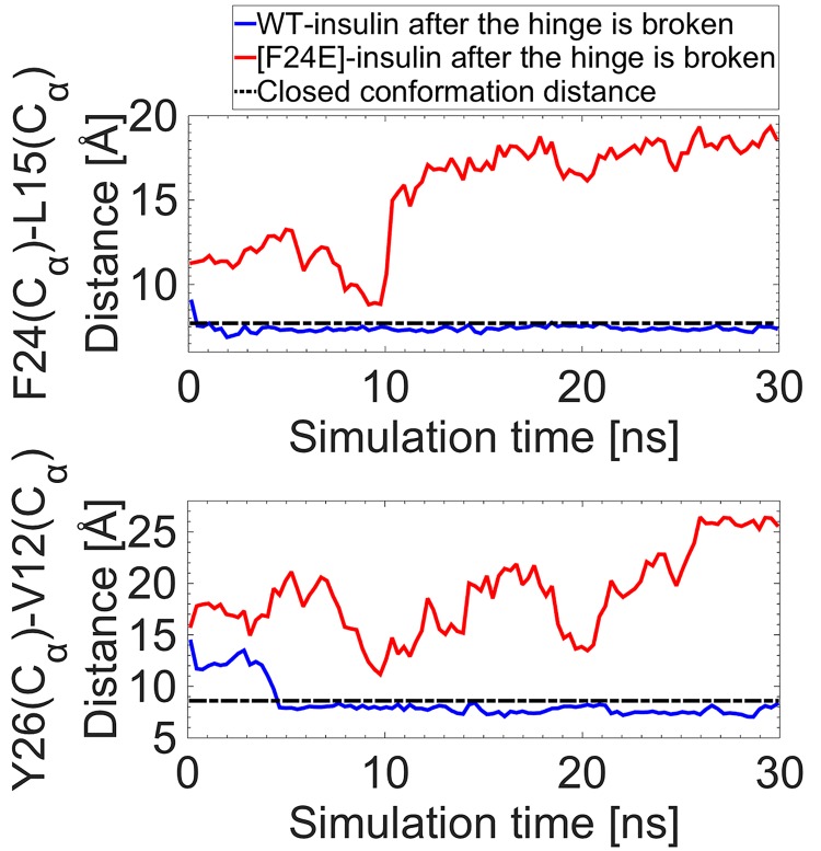
Fig 9. Stability of the hinge.
Distances between the Cα atoms of the F24–L15 and Y26–V12 pairs, which represent the hinge and the BC-CT opening, respectively, for the WT (blue) and the mutated (red) insulin, after the hinge is broken and the protein is in its wide-open conformation. The trajectories are compared to the closed conformation distance of WT insulin (black dashed line).

