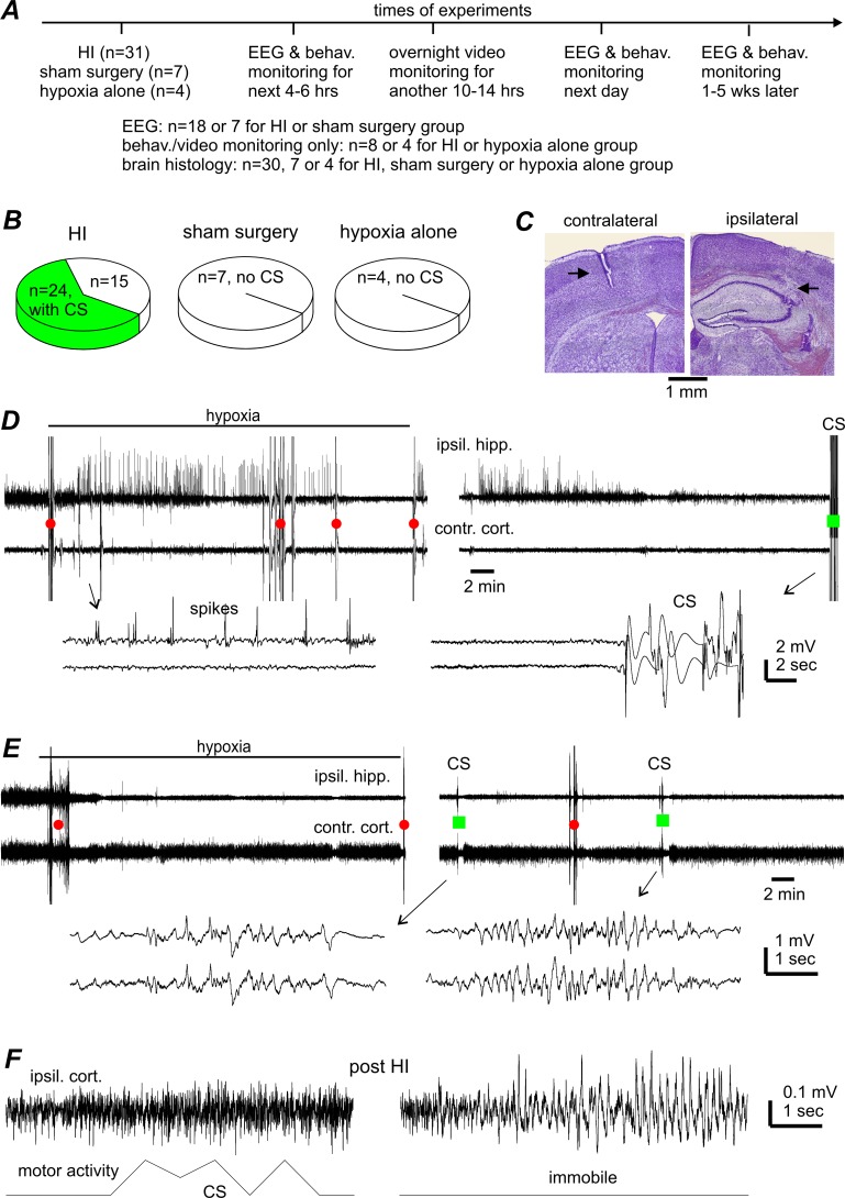
Fig 1. CS incidence and lack of CS-concurrent hippocampal-cortical EEG discharges.
A, a schematic presentation of experimental manipulations and animal groups. B, CS incidences in aging mice following HI, sham surgery or hypoxia alone. C, representative brain histologic sections obtained from one aging mouse, showing the tracks (black arrows) of implanted EEG electrodes in the ipsilateral hippocampus (right) and contralateral cortex (left). Similar observations were made in other 9 animals to confirm the location of implanted EEG electrodes. D-E, representative EEG traces were collected from 2 aging mice before, during, and shortly after hypoxia. Tethered recordings were made from the ipsilateral hippocampus (ipsil. hipp.) and contralateral parietal cortex (contra. cort.). Original data were treated with band-pass filtered (0.5–500 Hz) for illustration purpose. Red circles or green squares denote movement artifacts in the absence or presence of CS. Arrowed segments are expanded below, showing hippocampal spikes (D, left) and movement artifact-contaminated signals during CS (D, right and E, right). F, signals of ipsilateral cortical EEG (top) and gross motor activity (bottom) collected telemetrically from another aging mouse. Left, evident motor activity signals without corresponding EEG discharges during a CS event. Right, EEG spikes in the absence of motor activity signals (during immobility).

