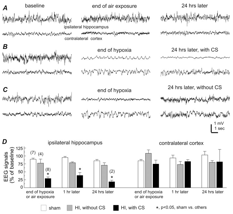
Fig 2. Decreases of ipsilateral EEG signals in aging mice with early-onset CS.
A-C, representative EEG traces collected from 3 aging mice during baseline monitoring (left), at the end of ambient air exposure or hypoxia (middle), and 24 hours later (right). Tethered recordings were made from the ipsilateral hippocampus and contralateral parietal cortex for each animal. Note the ipsilateral EEG suppression in the animal with post-HI CS (B), recovered ipsilateral EEG in the animal without CS (C) and the lack of EEG suppression in the control animal (A). D, 30-sec EEG segments were collected during baseline monitoring, at the end of either hypoxia or ambient air exposure, then at 1 hour, and 24 hours following either a sham operation or HI. The root mean square (RMS) of the EEG signals was calculated and normalized as a percentage of the baseline RMS. Animals are grouped as sham controls and post-HI with and without CS. Data (mean±SE) for the ipsilateral hippocampus (left) and the contralateral cortex (right) are presented separately. *, p<0.05, sham control vs. others, one way ANOVA.

