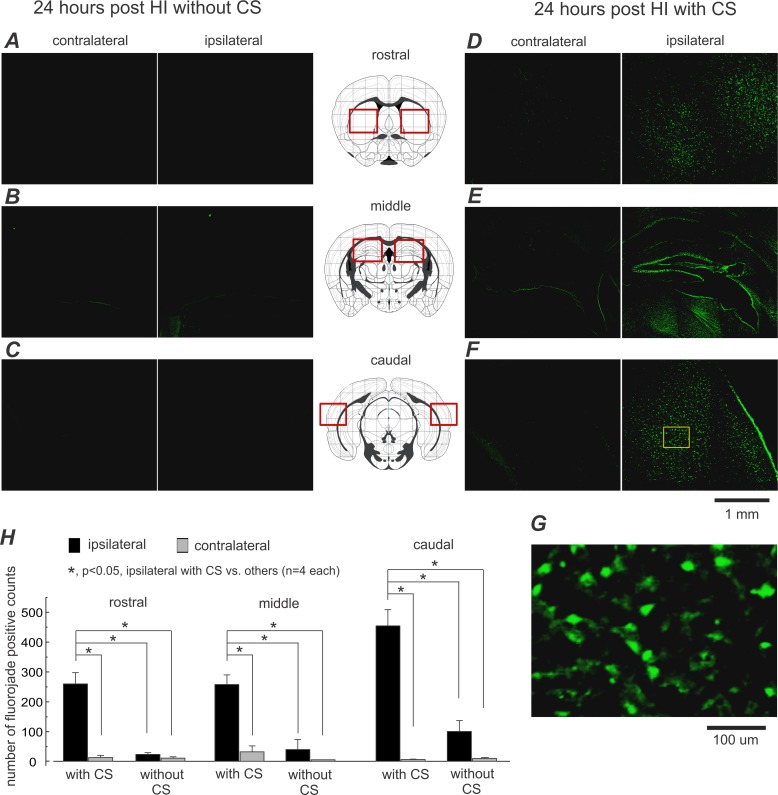
Fig 4. Abundant Fluoro-Jade positive cells in aging mice with post-HI CS.
A-F, representative images were collected from 2 aging mice 24 hours following HI, one with CS (D-F, right), the other without (A-C, left). Brain regions in which Fluoro-Jade positive cells were analyzed are indicated diagrammatically in middle column (by red squares). G, an enlarged image was taken from a selected cortical area (indicated by a yellow square in F). H, regional counts of Fluoro-Jade positive cells in 8 post-HI aging mice with and without CS (mean±SE; n = 4 in each group). *, p<0.05, ipsilateral cell counts in animals with CS vs. contralateral cell counts and cell counts in animals without CS, one way ANOVA.

