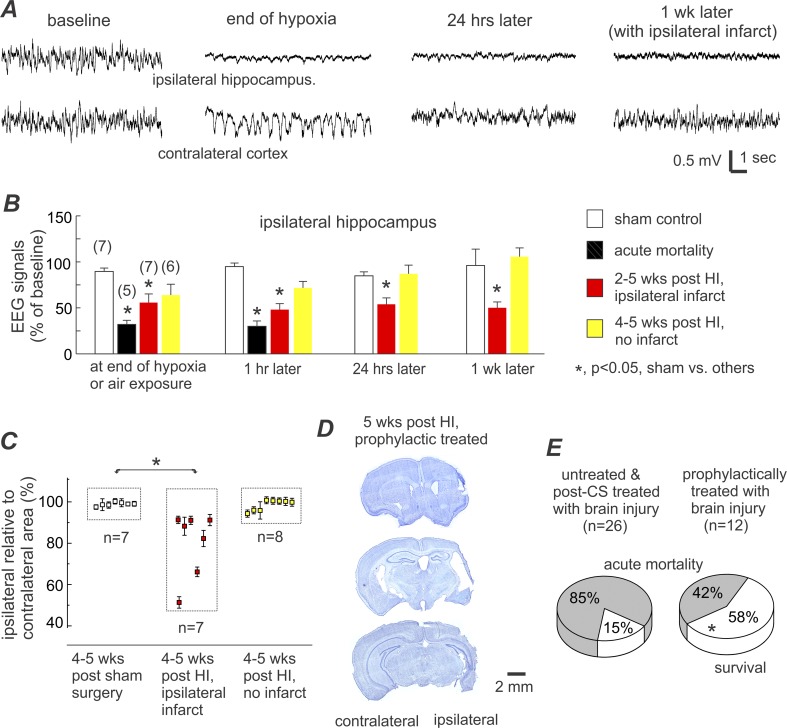
Fig 5. EEG and histological assessments and survival rate in prophylactic treated aging mice.
A, representative EEG traces were collected from a prophylactically treated aging mouse. Tethered recordings made from the ipsilateral hippocampus (top trace) and contralateral parietal cortex (bottom trace). Traces from left to right: baseline signals, at the end of hypoxia, 24 hours post-HI, and 1-week post-HI. B, quantification of EEG signals in sham control and prophylactically treated aging mice. Only the changes in the ipsilateral hippocampal signals are presented for brevity. Numbers of animals examined in different time points are indicated in parentheses. *, p<0.05, sham controls vs. other experimental groups, one-way ANOVA. C, ratios of ipsilateral relative to contralateral hemisphere areas were measured at 8 coronal levels and averaged for each animal. Data (%, mean±SE) for individual animals were pooled together according to the indicated experimental groups. D, representative images of brain sections were obtained from another prophylactically treated aging mouse. Histologic assessments were conducted 5 weeks post-HI, showing ipsilateral infarctions and decreased ipsilateral hemispheric area at three coronal levels. E, survival rates in two groups of aging mice under the indicated experimental conditions. Acute mortality was defined as spontaneous death or mandatory euthanization within 48 hours post-HI, and survival referred to animals that lived 4–5 weeks post-HI in acceptable physical condition. *, p = 0.026, prophylactically treated vs. untreated/post-CS treated, Fisher exact test.

