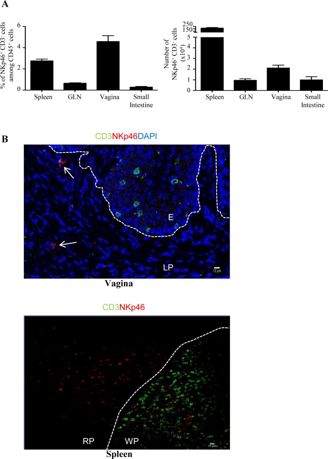
Fig 1. Identification of vaginal NKp46+ CD3- cells.
(A) Frequencies (left panel) and numbers (right panel) of NKp46+ CD3- cells among CD45+ leukocytes from spleen, genital lymph nodes (GLN) and vagina of naive C57BL/6 mice. Histogram plots represent results from five independent experiments and are expressed as mean values + SEM, n = 10 mice. (B) Immunofluorescence staining of frozen sections from mouse vagina and spleen stained with anti-CD3 (green) and anti-NKp46 (red) antibodies. Nuclei were visualized with DAPI (blue). White arrows indicate NKp46+ CD3- cells; E: epithelium; LP: lamina propria. WP: white pulp; RP: red pulp. White dotted lines delineate the epithelium from the lamina propria in the vagina and the white pulp from the red pulp in the spleen. Original magnification: x40 (vagina) and x20 (spleen). Data are representative of three independent experiments.

