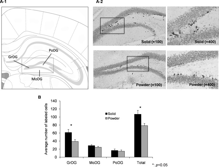
Fig 2. Neurogenesis in hippocampal DG.
(A-1) A schematic representation in the hippocampal DG area. GrDG: granule cell layer of DG, PoDG: polymorph layer of DG, MoDG: molecular layer of the DG. (A-2) Representative photomicrographs of BrdU labeled cells in hippocampal DG at one day after the last injection of BrdU in SD (top, N = 8) and PD (bottom, N = 8) group. (B) The total number and averaged number in each area of labeled cells are presented as mean ± SEM.

