
Figure 1.
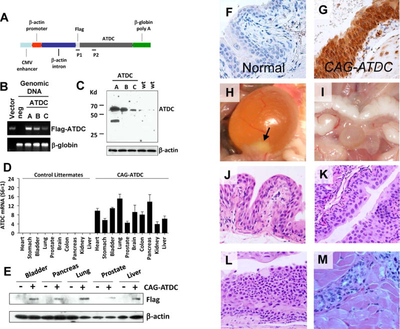
CAG-ATDC tg mice develop urothelial tumors similar to human tumors. (A) Diagrammatic illustration of the pCAGGS-flag-ATDC expression vector (CAG-ATDC). P1 and P2 indicate locations of primers for genotyping PCR. (B) Genotyping by qPCR of three CAG-ATDC transgenic mouse lines (A, B, and C) and an age-matched flag-ATDC negative littermate. pCAGGS-Flag-ATDC expression vector (Vector) was used as a positive control. β-globin was used as a PCR loading control. (C) Whole-cell lysates from the bladders of transgenic line A, B and C or control littermates were subjected to western blotting, and probed with ATDC antibody confirming ATDC tg expression. β-actin served as loading control. (D) Quantitative RT-PCR of flag-ATDC mRNA detected transgene expression in a wide variety of tissues in CAG-ATDC line A mice, but not in control littermates (n=6, mean±SE). (E) Western blotting of ATDC in protein lysates from various tissues confirms transgene protein expression in multiple tissues. (F) Immunostaining for ATDC in normal bladder littermate urothelium lacking transgene. (G) Immunostaining for ATDC in CAG-ATDC urothelium demonstrates high levels of expression. (H) A representative image of a bladder tumor (arrow) in CAG-ATDC mice (10 months of age). (I) A representative image of a normal bladder in a control littermate. (J) Representative H&E-stained sections depicting normal bladder urothelial histology in a control littermate (10 months of age). (K–M) Representative H&E-stained sections of CAG-ATDC lesions at 8–12 months of age showing low-grade noninvasive (K), carcinoma in situ (CIS) (L), and muscle-invasive (M) urothelial tumors.