
Figure 2.
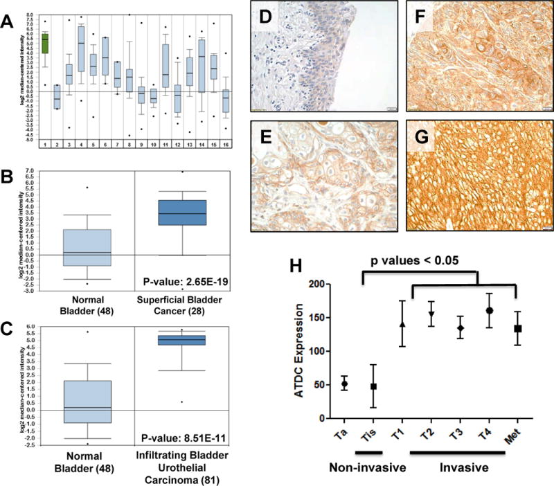
ATDC is highly expressed in human bladder cancers and correlates with invasive disease. (A) Comparison of ATDC expression in various tumor types in gene expression array data derived from Oncomine demonstrated that ATDC was most highly expressed in bladder tumors (p value = 2.98 × 10⁻¹²). 1: Bladder, 2: CNS, 3: Breast, 4: Cervical, 5: Colon, 6: Esophagus, 7: Gastric, 8: Head and Neck, 9: Kidney, 10: Liver, 11: Lung, 12: Lymphoma, 13: Ovarian, 14: Pancreatic, 15: Prostate, 16: Sarcoma. (B) Comparison of ATDC expression in normal human bladder and superficial human bladder tumors in Oncomine database demonstrated that ATDC was up-regulated in superficial tumors (p value =2.65×10⁻¹⁹) (30). (C) Comparison of ATDC expression in normal bladder and infiltrating (invasive) human bladder tumors demonstrated significant up-regulation of ATDC in tumors (p value = 8.51×10⁻¹¹) (30). For A–C, error bars represent 95% confidence intervals. (D–G) Representative images demonstrating absent ATDC expression in normal human urothelium (D) and low (E), moderate (F) or strong (G) expression in human invasive bladder tumor specimens. (H) Quantification of ATDC expression in non-invasive (n = 27) and invasive (n = 153) human urothelial tumors demonstrated higher ATDC expression in invasive human tumors (mean±SE). Each specimen was given a composite score (0–300) by a blinded bladder pathologist.