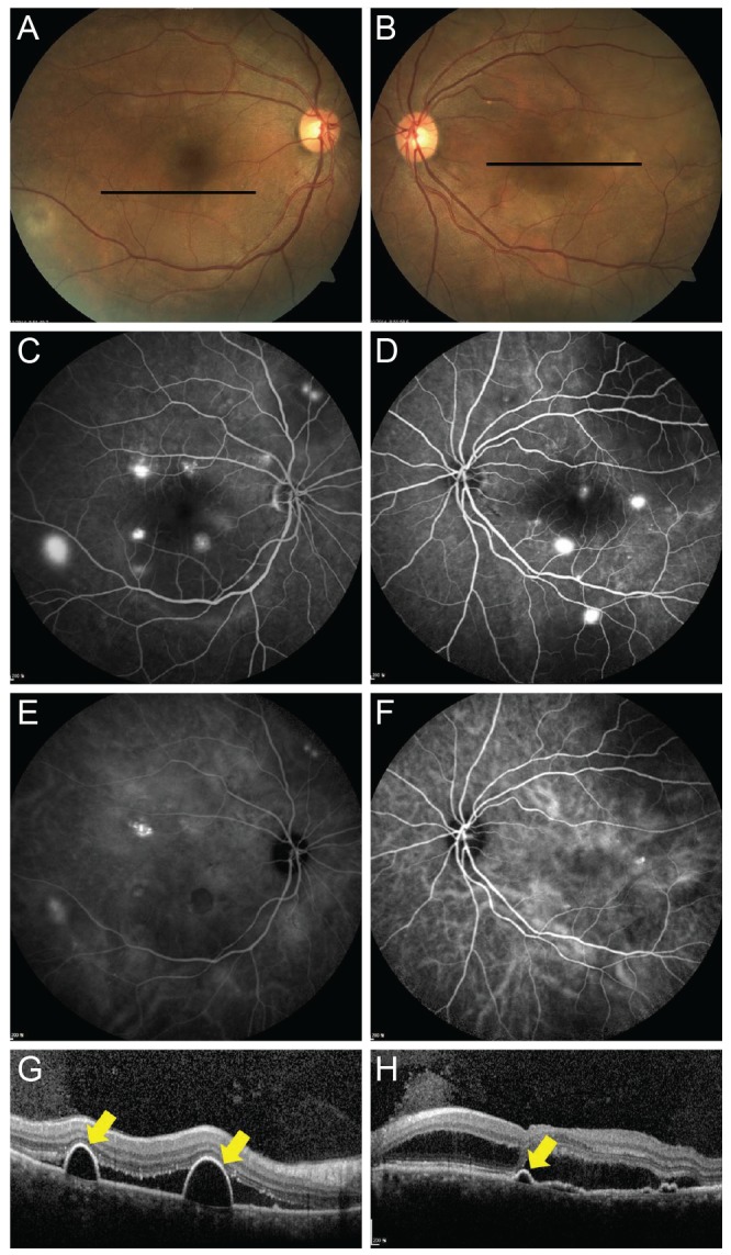
Fig. 2. Representative case of acute bilateral central serous chorioretinopathy. (A,B) On fundus examination, bilateral serous submacular fluids are observed. (C,D) Late-phase fluorescence angiography shows multifocal hyperfluorescent dots throughout posterior poles in both eyes. (E,F) Late-phase indocyanine green angiography shows multifocal hyperf luorescence, suggesting choroidal vascular hyperpermeability. (G,H) Optical coherence tomography (OCT) reveals subretinal fluid and thickened choroids. Additionally, OCT scans reveal pigment epithelial detachments (yellow arrows), corresponding to black lines in (A) and (B).

