. 2015 Nov 25;29(6):424–432. doi: 10.3341/kjo.2015.29.6.424

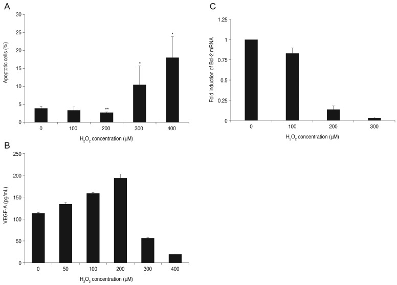
Fig. 2. Apoptosis and expression of vascular endothelial growth factor (VEGF)-A and B-cell leukemia/lymphoma (Bcl)-2 after treatment of retinal pigment epithelial (RPE) cells with H2O2. (A) Effects of H2O2 on apoptosis of RPE cells. The RPE cells were treated with various concentrations of H2O2 for 16 hours. Cell apoptosis was 3.26% for RPE at 100 µM H2O2 compared to 3.82% for the control, but the difference was not significant. Cell apoptosis decreased at 200 µM H2O2 (2.65%) but increased at 300 and 400 µM of H2O2 (10.42%, 17.99%). Means were significantly different than the control (p < 0.05). Each bar shows the mean ± standard deviation of results of three or more independent experiments. The asterisk indicates a statistically significant difference within the group (*increased, **decreased, p < 0.05). (B) Expression of VEGF-A after exposure to H2O2. VEGF-A excretion into the medium was measured using enzyme-linked immunosorbent assay. VEGF-A expression increased after addition of 50, 100, or 200 µM H2O2 to RPE cells. However, after being treated with 300 and 400 µM H2O2, VEGF-A expression decreased. Data are expressed as the mean ± standard deviation of the results of three or more independent experiments. Means were statistically significant compared to the control at all concentrations of H2O2 (p < 0.05). (C) Expression of Bcl-2 mRNA after exposure to H2O2. Cells were cultured with various concentrations of H2O2 for 16 hours (0, 100, 200, and 300 µM). Expression of Bcl-2 mRNA decreased as oxidative stress increased. Data are expressed as the mean ± standard deviation of the results of three or more independent experiments. Means were statistically significant compared with the control at all concentrations of H2O2 (p < 0.05).

Fig. 2