. 2015 Nov 25;29(6):424–432. doi: 10.3341/kjo.2015.29.6.424

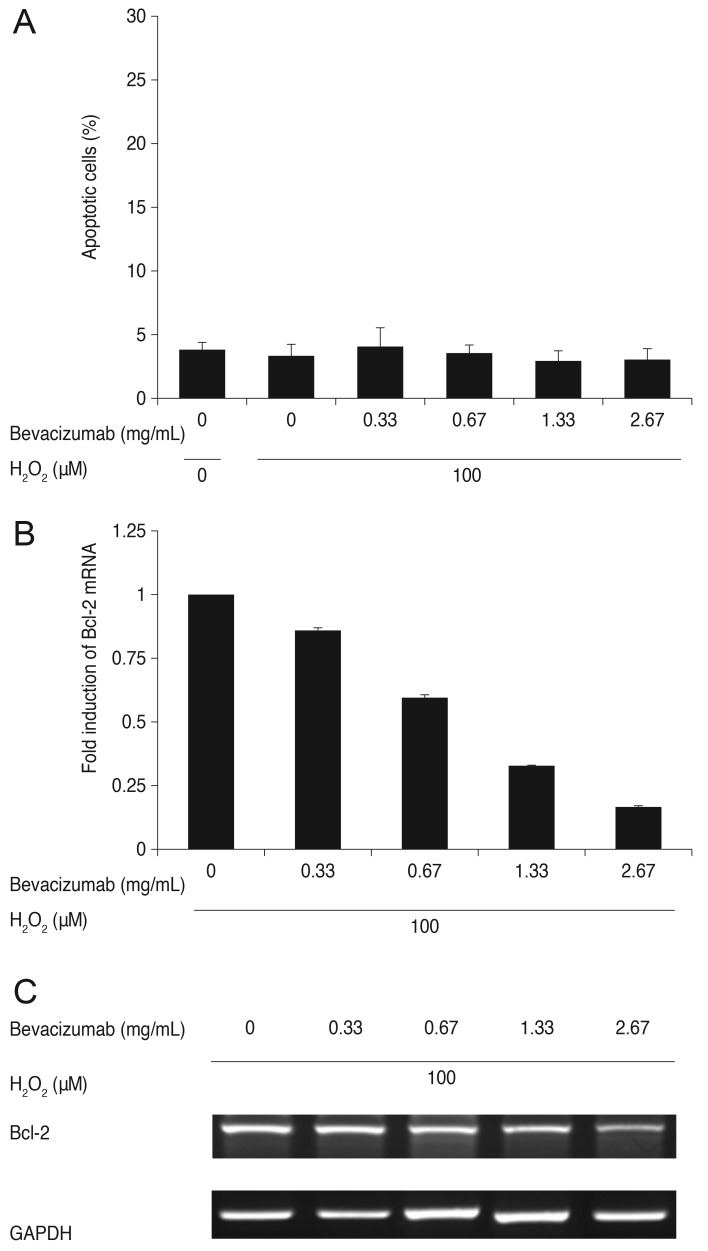
Fig. 3. Influence of bevacizumab on apoptosis of retinal pigment epithelial (RPE) cells and B-cell leukemia/lymphoma (Bcl)-2 mRNA expression under low oxidative stress (100 µM H2O2). (A) Influence of bevacizumab on apoptosis of RPE cells under low oxidative stress (100 µM H2O2). The RPE cells were treated with various concentrations of bevacizumab (0, 0.33, 0.67, 1.33, and 2.67 mg/mL) under low oxidative stress (100 µM H2O2). Cell apoptosis was not significantly different at all concentrations of bevacizumab less than 100 µM H2O2. Each bar shows the mean ± standard deviation of results of three or more independent experiments. p < 0.05 vs. control. (B,C) Expression of Bcl-2 mRNA with bevacizumab under low oxidative stress (100 µM H2O2). Expression of Bcl-2 mRNA decreased under low oxidative stress (100 µM H2O2), and this decrease was proportional to the increase in bevacizumab dose. There was a statistically significant difference at all concentrations. The mRNA expression of Bcl-2 was normalized to glycerol-3-phosphate dehydrogenase (GAPDH, housekeeping gene). Each bar shows the mean ± standard deviation of results of three or more independent experiments. p < 0.05 vs. control.

Fig. 3