Figure 5.
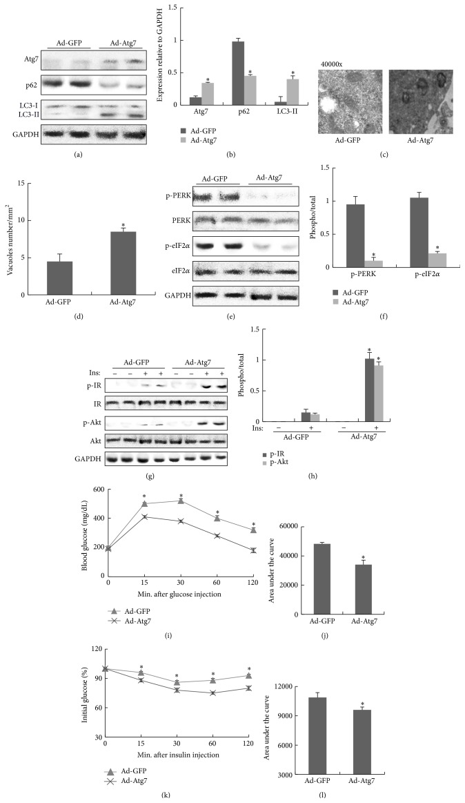
Improvement of ER stress and insulin signaling by the restoration of Atg7 in liver of obese mice. All analyses were performed in mice fed a high-fat diet (HFD) for 8 weeks and then adenovirus carrying Atg7 or GFP was delivered into obese mice via orbital venous plexus at a titer of 3 × 10¹¹ vp/mice. (a) Protein expression of Atg7, p62, and LC3 in liver. (b) The relative protein quantity of Atg7, p62, and LC3 in liver. (c) Quantification of autophagolysosome-like vacuoles per field in the EM images of liver (magnification 40000x). (d) Quantification of autophagolysosome-like vacuoles per field in the EM images of liver. (e) Phosphorylation of PERK and eIF2α in liver. (f) The relative protein quantity of p-PERK and p-eIF2α in liver. (g) Phosphorylation of IR and Akt in liver. (h) The relative protein quantity of p-IR and p-Akt in liver. (i) GTT. (j) Area under the curve by GTT. (k) ITT. (l) Area under the curve by ITT. The relative quantity of proteins was analyzed using Quantity One software. A representative blot is shown and the data was expressed as mean ± SEM in each bar graph. ∗ P < 0.05 (HFD/Ad-Atg7 versus HFD/Ad-GFP).

