
Figure 1. The detection limit for exogenous RML infection.
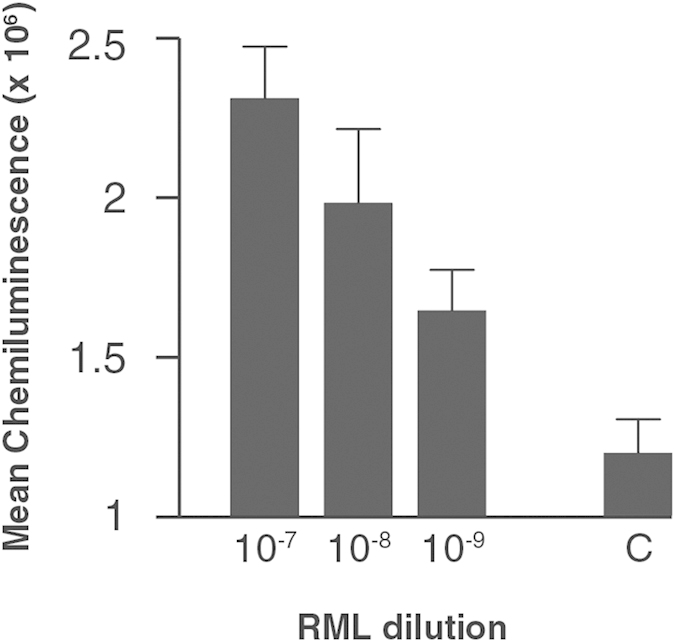
A 10% w/v RML-prion infected brain homogenate was logarithmically diluted into whole blood collected from wild-type CD-1 mice down to 10−9 and analysed by DDA. Mean chemiluminescence signals with standard errors of the mean (SEM) are shown for two independent experiments with n = 3 for each experiment. The signal arising from the whole blood diluent alone is shown as a background control (C).