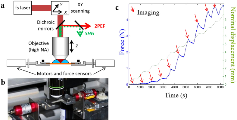
Figure 1. Experimental setup and protocol.
(a) Scheme of the combined multiphoton microscope and traction device. 2PEF and SHG are detected in the backward direction; (b) View of the skin sample maintained under the microscope objective by two jaws fixed to two motors and two force sensors; (c) Experimental timetable showing stepwise increasing stretching of the skin sample (green dotted line) with continuous force measurement (blue straight line) and time-lapse imaging of immobile sample after each displacement step (red arrows).

