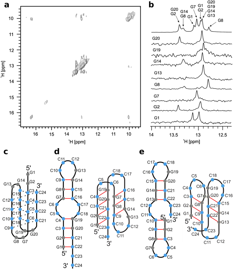
Figure 3. Assignment of guanine imino protons and proposed structures.
(a) Imino-imino region of NOESY spectrum of d(G2C4)4 at mixing time of 150 ms. (b) Assignment of imino protons of guanine residues involved in GC base pairs using 1D 15N-edited HSQC NMR spectra of residue specific (marked on the left side of each spectrum) 15N, 13C-isotopically labelled d(G2C4)4. Top spectrum in B represents imino region of 1H NMR spectrum. All spectra were acquired at pH 4.7 and 5 °C. (c) Proposed structures of i-motif and (d,e) hairpins adopted by d(G2C4)4.

