
Figure 1.
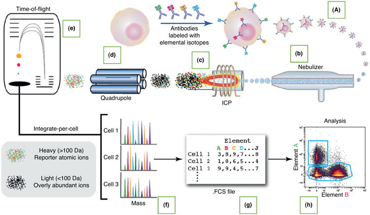
Workflow of mass cytometry analysis. A liquid sample containing cells labeled with heavy metal isotope-conjugated probes (ICPs) (a) is introduced into the nebulizer (b), where it is aerosolized. The aerosol droplets are directed into the ICP torch (c), where the cells are vaporized, atomized, and ionized. Low-mass ions are removed in the radiofrequency (RF) quadrupole ion guide (d), resulting in a cloud of ions enriched for the probe isotopes. The ion cloud then enters the time-of-flight (TOF) chamber (e), where the ions are separated on the basis of their mass:charge ratio as they accelerate toward the detector. Thus, the time-resolved detector measures a mass spectrum (f) that represents the identity and quantity of each isotope on a per cell basis. Data are generated in .fcs format (g) and analyzed using the cloud-based Cytobank platform (h). Reproduced from [40].