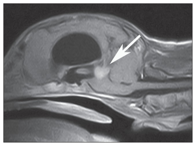
Figure 1.
Postcontrast midsagittal SE T1-weighted magnetic resonance image of the brain of dog 1. Notice the homogeneous enhancement of the rounded mass occupying the dorsal region of the mesencephalon (arrow). Severe dilation of the lateral and third ventricles is evident. A marked mass effect, displacing and compressing ventrally the brainstem and caudally the cerebellum, is evident. (Reprinted from: Thalamic astrocytic hamartoma and associated meningoangiomatosis in a German shepherd dog. In: Pasquali P, ed. Research in Veterinary Science. Vol 94. Philadelphia, Pennsylvania: Elsevier, 2013:644–647, with permission).

