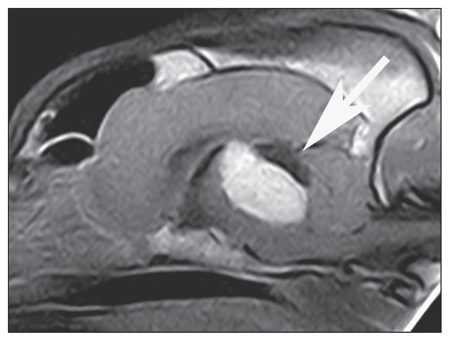
Figure 2.
Postcontrast midsagittal SE T1-weighted magnetic resonance image of the brain of dog 2. Notice the homogeneous enhancement of the ovoid mass located at the level of the dorsal portion of mesencephalon (arrow). The mass extends from the level of the thalamus to the level of the medulla. A marked mass effect is evident, displacing and compressing the cerebellum caudally, the brainstem ventrally, and the diencephalic structures cranially.

