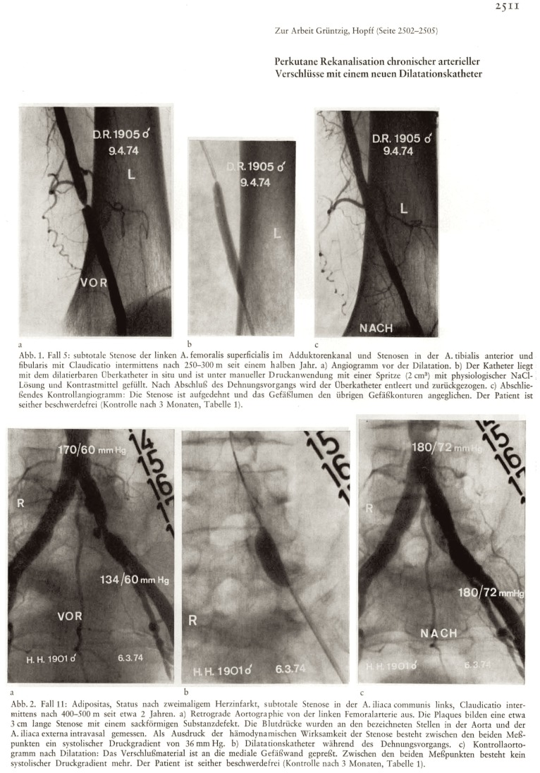
Figure 3.
Supplemental information (“Zur Arbeit Grüntzig, Hopff”) to the article depicted in Figure 2, showing arterial angiograms of two patients with peripheral artery disease before (left), during (middle), and after (right) percutaneous balloon angioplasty performed by Andreas Grüntzig, M.D. The first angiogram in panel 1 (Abb. 1) indicates left superficial femoral artery stenosis in a 71-year-old man, the procedure was performed on April 9, 1974. The left angiogram panel 2 (Abb. 2) indicates a high-grade stenosis of the left common iliac artery in a 73-year-old male patient, the procedure was performed on March 6, 1974. Note the differently sized angioplasty balloons, both in diameter and length. The German figure legend indicates that both patients were free of symptoms on follow-up 3 months after the procedure. Reproduced from Grüntzig and Hopff (10), with permission of the publisher.

