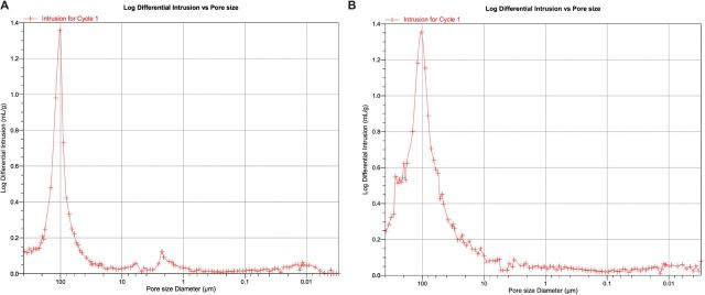
Figure 5.
(A and B) Representative images of control scaffold with no nanofibers (A-left) and 0.25% hybrid scaffold (B-right). Although differences are notable between the control and hybrid nanofiber preparations, most importantly both scaffolds maintain a pore distribution that includes abundant pores around 100 µm in diameter. It is believed that this is the lower limit of pores that are conducive to bone tissue formation.

