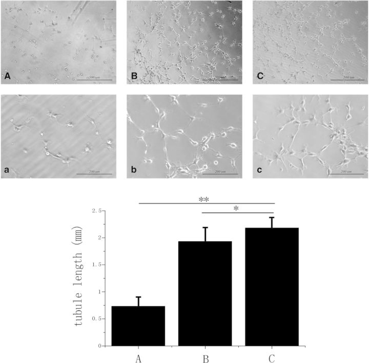
Fig. 3.
The in vitro tube formation assay of EPCs. (A) Without coating, scale bars = 500 µm; (a): without coating, scale bars = 200 µm. (B) Anti-CD34 antibody coating, scale bars = 500 µm; (b): anti-CD34 antibody coating, scale bars = 200 µm. (C) Anti-CD133 antibody coating, scale bars = 500 µm; (c): anti-CD133 antibody, scale bars = 200 µm (*P < 0.05; **P < 0.01)

