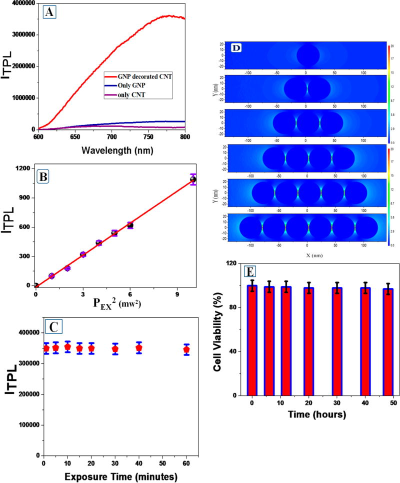
Figure 2.

(A) Two-photon photoluminescence spectrum from anti-GD2 antibody conjugated SWCNT–GNP theranostic material, anti-GD2 antibody conjugated GNP, and anti-GD2 antibody conjugated SWCNT. NIR light (1100 nm) was used as the excitation source. (B) Plot indicates linear relationship between two-photon photoluminescence intensity at 775 nm from anti-GD2 antibody conjugated SWCNT–GNP theranostic material and the square of intensity of 1100 nm excitation laser power (PEX). (C) Plot indicates two-photon luminescence at 775 nm from anti-GD2 antibody conjugated SWCNT–GNP remain same even after an hour of exposure with 1100 nm light. (D) Plot reports FDTD simulated electric field enhancement |E|2 profiles (arb. unit) for nanoparticle assembly. For our calculation, we have used GNPs particle size as 40 nm, and separation distance between GNPs are kept as 3 nm. (E) Bar plot indicates very good biocompatibility of anti-GD2 antibody conjugated SWCNT–GNP theranostic against melanoma UACC903 cells. Even after 48 h incubation with 80 μg of hybrid material, we have observed about 97% cell viability.
