
Fig. 3. Effect of CSP and XIP on cell survival of S. mutans.
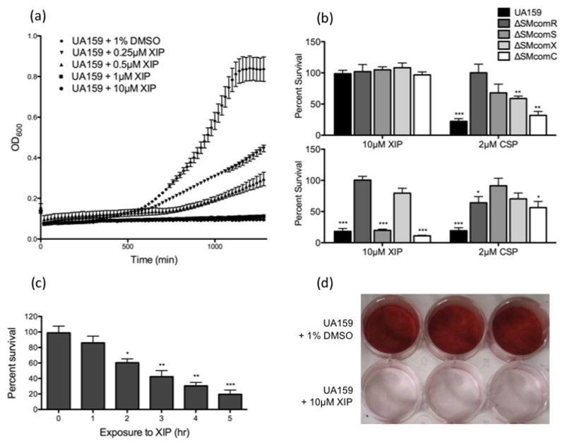
(a). The effect of XIP at varying concentrations (10μM, 1μM, 0.5μM, 0.25μM, 1% DMSO) on the growth of S. mutans UA159 was examined by monitoring OD600 using the Bioscreen C automated growth reader (b). S. mutans UA159, as well as comR, comS, comX and comC knockout mutant strains were grown in the presence or absence of either XIP (10μM) or CSP (2μM) to OD600~0.8 in THYE (top panel) or CDM (bottom panel). Viable CFUs were counted and results were standardized to the dry weight and presented as the percentage of the CFUs obtained in the absence of the peptide. Results shown here are the average of four independent experiments ± standard error. Statistical analyses were performed using Student’s T-test: *p<0.05, **p<0.01, ***p<0.001. (c). Actively growing cultures of UA159 were exposed to 10μM XIP for 0, 1, 2, 3, 4 and 5h. Following sonication, viable CFUs were counted and results were standardized to the dry weight and presented as the percentage of the CFUs obtained in the absence of the peptide. Results shown here are the average of four independent experiments ± standard error. Statistical analyses were performed using Student’s T-test: *p<0.05, **p<0.01, ***p<0.001. (d). S. mutans biofilms were grown on polystyrene plates overnight in the presence or absence of 10μM XIP, and stained with 0.1% Safranin Red. The image presented here is a representative of four independent experiments.