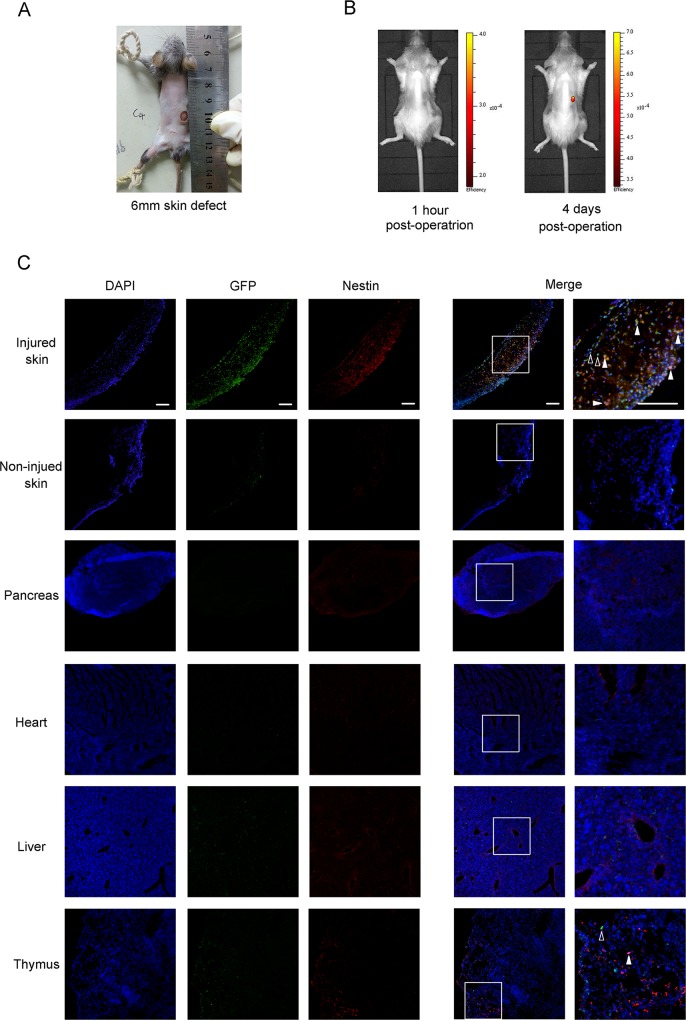
Fig 2. Nestin+ BMDCs are mobilized to the skin injury position.
(A) General view of the just made 6mm full thickness skin defect. (B) In vivo fluorescence imaging of GFP+ BMDCs distribution 1 hour or 4 days after the skin defect made. (C) Immunofluorescence GFP+/nestin+ double positive cells around injured skin and other organs 4 days after operation. Solid arrows indicate the GFP+/nestin+ double positive cells and hollow arrows indicate GFP single positive cells. All scale bars, 100μm.

