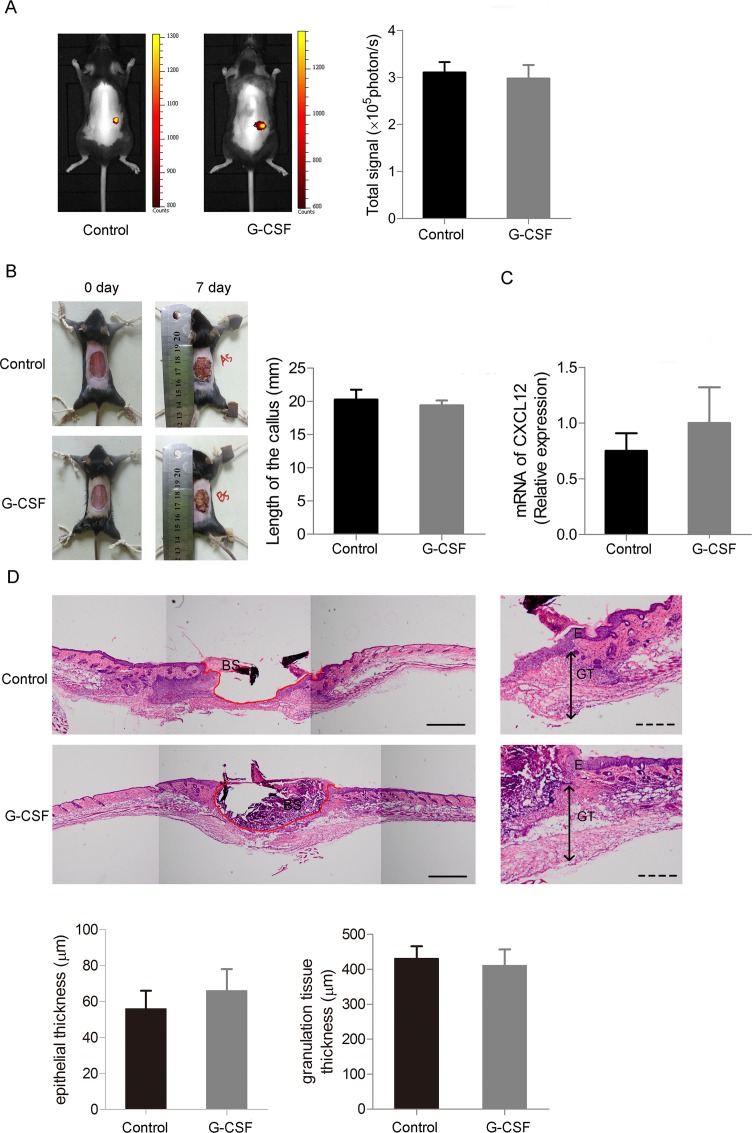
Fig 3. G-CSF does not promote mobilizing BMDCs and takes no effect on skin defect recovery.
(A) In vivo fluorescence imaging analysis of GFP+ BMDCs distribution 4 days after the skin defect made given G-CSF or normal saline. (B) Left: General view of the 20mm skin defect 7 days after the operation given G-CSF or normal saline. Right: Statistics of callus length of the two groups. (C) RT-PCR of CXCL12 of the skin tissue from the two groups of mice. (D) Re-epithelialization of the injured skin of the two groups of mice. Upper: Representative HE staining images showing marginal location of the injured skin tissues. (The red lines indicate the outlines of the skin defects. E: epidermis, GT: granulation tissue and BS: blood scab.) Lower: Statistics of epithelial and granulation tissue thickness. Continuous scale bars, 500μm. Dotted scale bars, 200μm. (n = 4 per group). Data (± SD) are representative of three independent experiments. Student’s t test was performed to determine statistical significance (* p<0.05).

