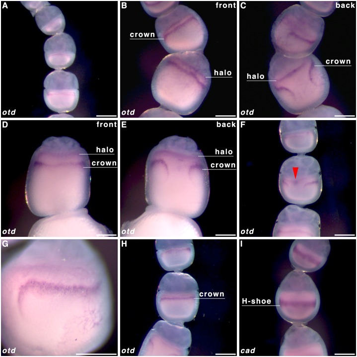
Fig 5. orthodenticle transcript localisation in Pararge aegeria oocytes.
Ovarioles were hybridised with a riboprobe targeting otd RNA (A-H). For comparison purposes cad localisation in an oocyte of a similar stage to H is also shown in panel I. Transcripts for otd localise to 2 distinct domains; an anterior domain (halo) lining the nurse cell-oocyte boundary and a more posterior domain (crown); a narrow band that breaks after curving posteriorly on one face. Panels C and E depict the back view of the ovarioles in panels B and D respectively. All ovarioles are oriented in such a way that the AP axis in maturing oocytes is depicted top to bottom (i.e. anterior of oocyte is bordering the nurse cells). Scale bars 200 μm.

