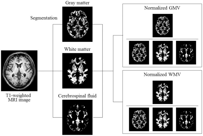
Fig 1. Segmentation of brain magnetic resonance imaging (MRI) and normalization of gray matter volume (GMV) and white matter volume (WMV).
Representative axial brain image of T1-weighted MRI and segmented images of gray matter, white matter, and cerebrospinal fluid are shown. To normalize for head size variability, GMV and WMV were normalized by dividing by the total intracranial volume, calculated by adding GMV, WMV, and cerebrospinal fluid space volume.

