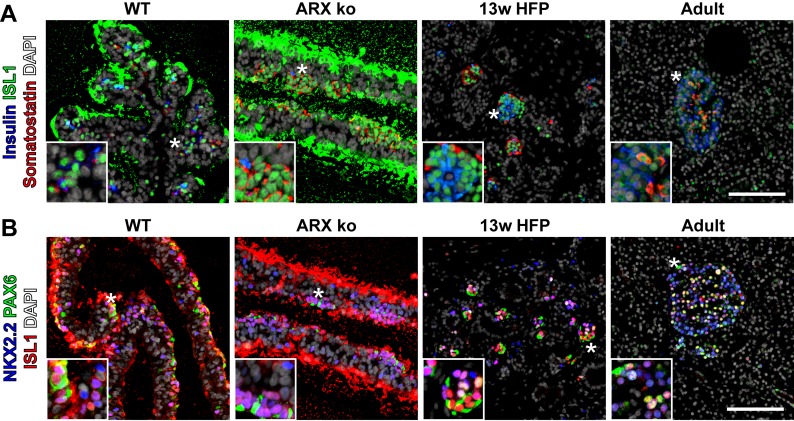
Fig 7. Expression of ISL1 in ARX ko hESCs and Human Tissue.
(A) Immunostaining of ISL1 (green), somatostatin (red), and insulin (blue) in 26 day differentiated wild type (WT) and ARX knockout cells (ARX ko), 13 week human fetal pancreatic tissue, and adult human pancreatic tissue. (B) Immunostaining of PAX6 (green), ISL1 (red), and NKX2.2 (blue) in the same sample series as above. Inset is a ~3x enlarged portion from the region indicated by the star. Scale bar is 100 μm for all panels. Extracellular immunoreactivitity of ISL1 is a staining artifact associated with the agarose which surrounds the embedded cell sheets.

