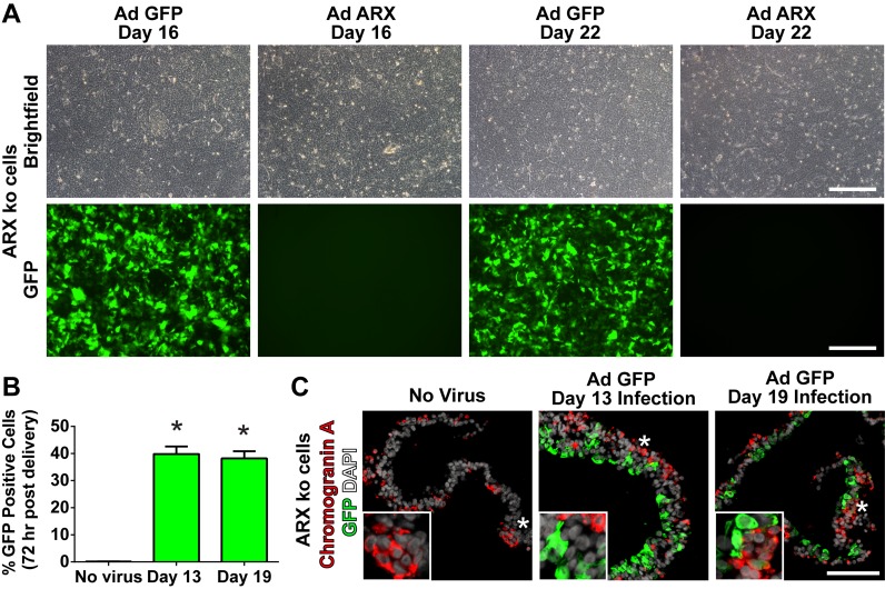
Fig 11. Adenoviral Infection Efficiency in ARX ko Cells.
(A) Brightfield and green channel (GFP) images of differentiating ARX knockout (ARX ko) cell cultures 72 hours post adenoviral infection at a MOI of 2. (B) GFP infection efficiency quantified by flow cytometry 72 hours after viral delivery as a percentage of the total population. (C) Transgene expression (GFP) in 26-day differentiated ARX ko hESCs based on immunostatining of GFP (green) and chromogranin A (red). Nuclei are counterstained with DAPI (white). Inset in C is a ~3x enlarged portion from the region indicated by the star. Scale bar is 200 μm for panel A and 100 μm for C.

