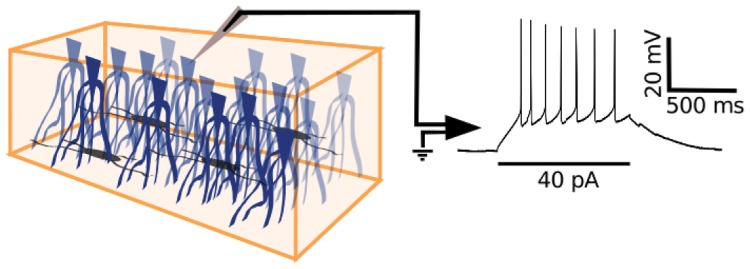
Fig 2. Whole-cell recordings from pyramidal neurons in turtle visual cortex.
Schematic diagram of an isolated piece of turtle dorsal cortex (left panel) with the ventricular side up and containing pyramidal neurons (blue) and interneurons (grey). A whole-cell recording of the pyramidal neuron membrane potential in response to current injection (right panel) is obtained with a patch electrode (grey triangle) that is positioned at the pyramidal neuron soma under visual guidance with DIC optics.

